
[ad_1]
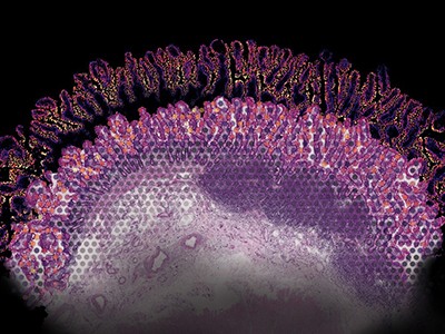
One cubic millimetre doesn’t sound like a lot. However within the human mind, that quantity of tissue comprises some 50,000 neural ‘wires’ linked by 134 million synapses. Jeff Lichtman wished to hint all of them.
To generate the uncooked information, he used a protocol referred to as serial thin-section electron microscopy, imaging 1000’s of slivers of tissue over 11 months. However the information set was monumental, amounting to 1.4 petabytes — the equal of about 2 million CD-ROMs — far an excessive amount of for researchers to deal with on their very own. “It’s merely not possible for human beings to manually hint out all of the wires,” says Lichtman, a molecular and cell biologist at Harvard College in Cambridge, Massachusetts. “There will not be sufficient individuals on Earth to actually get this job executed in an environment friendly means.”
It’s a typical chorus in connectomics — the examine of the mind’s structural and practical connections — in addition to in different biosciences, by which advances in microscopy are making a deluge of imaging information. However the place human sources fail, computer systems can step in, particularly deep studying algorithms which have been optimized to tease out patterns from massive information units.
“We’ve actually had a Cambrian explosion of instruments for deep studying previously few years,” says Beth Cimini, a computational biologist on the Broad Institute of MIT and Harvard in Cambridge, Massachusetts.
Deep studying is an artificial-intelligence (AI) method that depends on many-layered synthetic neural networks impressed by how neurons interconnect within the mind. Primarily based as they’re on black-box neural networks, the algorithms have their limitations. These embody a dependence on huge information units to show the community how one can determine options of curiosity, and a generally inscrutable means of producing outcomes. However a fast-growing array of open-source and web-based instruments is making it simpler than ever to get began (see ‘Taking the leap into deep studying’).
Listed below are 5 areas by which deep studying is having a deep affect in bioimage evaluation.
Massive-scale connectomics
Deep studying has enabled researchers to generate more and more advanced connectomes from fruit flies, mice and even people. Such information can assist neuroscientists to know how the mind works, and the way its construction modifications throughout growth and in illness. However neural connectivity isn’t simple to map.
In 2018, Lichtman joined forces with Viren Jain, head of Connectomics at Google in Mountain View, California, who was on the lookout for an appropriate problem for his staff’s AI algorithms.
“The picture evaluation duties in connectomics are very troublesome,” Jain says. “You’ve gotten to have the ability to hint these skinny wires, the axons and dendrites of a cell, throughout massive distances, and standard image-processing strategies made so many errors that they had been mainly ineffective for this job.” These wires may be thinner than a micrometre and prolong over lots of of micrometres and even millimetres of tissue. Deep-learning algorithms present a option to automate the evaluation of connectomics information whereas nonetheless reaching excessive accuracy.
Tips on how to make spatial maps of gene exercise — all the way down to the mobile stage
In deep studying, researchers can use annotated information units containing options of curiosity to coach advanced computational fashions in order that they will rapidly determine the identical options in different information. “Whenever you do deep studying, you say, ‘okay, I’ll simply give examples and you work every thing out’,” says Anna Kreshuk, a pc scientist on the European Molecular Biology Laboratory in Heidelberg, Germany.
However even utilizing deep studying, Lichtman and Jain had a herculean job in making an attempt to map their snippet of the human cortex1. It took 326 days simply to picture the 5,000 or so extraordinarily skinny sections of tissue. Two researchers spent about 100 hours manually annotating the photographs and tracing neurons to create ‘floor fact’ information units to coach the algorithms, in an method referred to as supervised machine studying. The educated algorithms then routinely stitched the photographs collectively and recognized neurons and synapses to generate the ultimate connectome.
Jain’s staff introduced huge computational sources to bear on the issue, together with 1000’s of tensor processing items (TPUs), Google’s in-house equal to graphics processing items (GPUs) constructed particularly for neural-network machine studying. Processing the info required on the order of 1 million TPU hours over a number of months, Jain says, after which human volunteers proofread and corrected the connectome in a collaborative course of, “type of like Google Docs”, says Lichtman.
The tip outcome, they are saying, is the biggest such information set reconstructed at this stage of element in any species. Nonetheless, it represents simply 0.0001% of the human mind. However as algorithms and {hardware} enhance, researchers ought to have the ability to map ever bigger parts of the mind, whereas having the decision to identify extra mobile options, corresponding to organelles and even proteins. “In some methods,” says Jain, “we’re simply scratching the floor of what is likely to be attainable to extract from these photos.”
Digital histology
Histology is a key device in medication, and is used to diagnose illness on the idea of chemical or molecular staining. Nevertheless it’s laborious, and the method can take days and even weeks to finish. Biopsies are sliced into skinny sections and stained to disclose mobile and sub-cellular options. A pathologist then reads the slides and interprets the outcomes. Aydogan Ozcan reckoned he may speed up the method.
Python power-up: new picture device visualizes advanced information
{An electrical} and laptop engineer on the College of California, Los Angeles, Ozcan educated a customized deep-learning mannequin to stain a tissue part computationally by presenting it with tens of 1000’s of examples of each unstained and stained variations of the identical part, and letting the mannequin work out how they differed.
Digital staining is nearly instantaneous, and board-certified pathologists discovered it virtually not possible to differentiate the ensuing photos from conventionally stained ones2. Ozcan has additionally proven that the algorithm can replicate a molecular stain for the breast most cancers biomarker HER2 in seconds, a course of that usually takes a minimum of 24 hours in a histology lab. A panel of three board-certified breast pathologists rated the photographs as having comparable high quality and accuracy to standard immunohistochemical staining3.
Ozcan, who goals to commercialize digital staining, hopes to see functions in drug growth. However by eliminating the necessity for poisonous dyes and costly staining tools, the method may additionally enhance entry to histology companies worldwide, he says.
Cell discovering
If you wish to extract information from mobile photos, you need to know the place within the photos the cells really are.
Researchers often carry out this course of, referred to as cell segmentation, both by cells beneath the microscope or outlining them in software program, picture by picture. “The phrase that almost all describes what individuals have been doing is ‘painstaking’,” says Morgan Schwartz, a computational biologist on the California Institute of Expertise in Pasadena, who’s growing deep-learning instruments for bioimage evaluation. However these painstaking approaches are hitting a wall as imaging information units develop into ever bigger. “A few of these experiments you simply couldn’t analyse with out automating the method.”
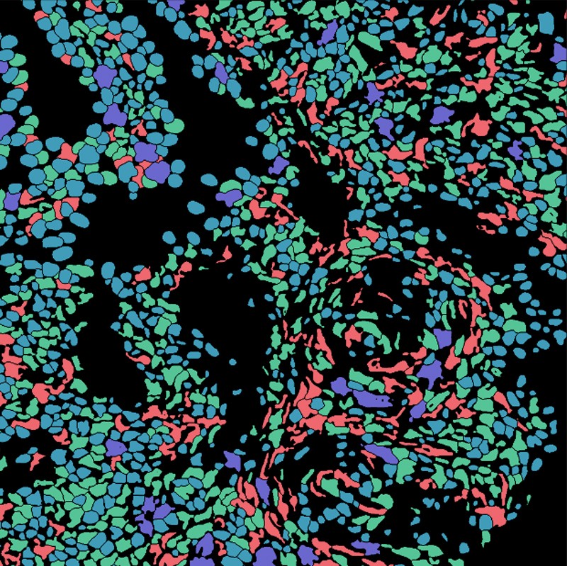
Lineage-based segmentation reveals the form of cells within the lining of the uterus throughout human being pregnant.Credit score: N. F. Greenwald et al. Nature Biotechnol. 40, 555–565 (2022).
Schwartz’s graduate adviser, bioengineer David Van Valen, has created a set of AI fashions, accessible at deepcell.org, to rely and analyse cells and different options from photos each of reside cells and of preserved tissue. Working with collaborators together with Noah Greenwald, a most cancers biologist at Stanford College in California, Van Valen developed a deep-learning mannequin referred to as Mesmer to rapidly and precisely detect cells and nuclei throughout totally different tissue sorts4. “In the event you’ve obtained information that you just want processed, now you’ll be able to simply add them, obtain the outcomes and visualize them both throughout the net portal or utilizing different software program packages,” Van Valen says.
In keeping with Greenwald, researchers can use such data to distinguish cancerous from non-cancerous tissue and to seek for variations earlier than and after therapy. “You’ll be able to take a look at the imaging-based modifications to have a greater thought of why some sufferers reply or don’t reply, or to determine subtypes of tumours,” he says.
Mapping protein localization
The Human Protein Atlas undertaking exploits yet one more software of deep studying: intracellular localization. “We’ve got for many years been producing tens of millions of photos, outlining the protein expression in cells and tissues of the human physique,” says Emma Lundberg, a bioengineer at Stanford College and a co-manager of the undertaking. At first, the undertaking annotated these photos manually. However as a result of that method wasn’t sustainable long run, Lundberg turned to AI.

NatureTech hub
Lundberg first mixed deep studying with citizen science, tasking volunteers with annotating tens of millions of photos whereas taking part in a massively multiplayer sport, EVE On-line5. Over the previous few years, she has switched to a crowdsourced AI-only resolution, launching Kaggle challenges — by which scientists and AI lovers compete to realize numerous computational duties — of US$37,000 and $25,000, to plot supervised machine-learning fashions to annotate protein-atlas photos. “The Kaggle problem afterwards blew the avid gamers away,” Lundberg says. The profitable fashions outperformed Lundberg’s earlier efforts at multi-label classification of protein-localization patterns by about 20% and had been generalizable throughout cell strains6. And so they managed one thing no printed fashions had executed earlier than, she provides, which was to precisely classify proteins that exist in a number of mobile areas.
“We’ve got proven that half of all human proteins localized to a number of mobile compartments,” says Lundberg. And placement issues, as a result of the identical protein would possibly behave in a different way elsewhere. “Realizing if a protein is within the nucleus or within the mitochondria, it helps perceive plenty of issues about its perform,” she says.
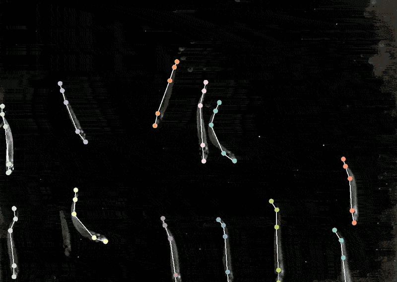
Annotation of fish for DeepLabCut coaching.Credit score: J. Lauer et al. Nature Strategies 19, 496–504 (2022). (CC BY 4.0)
Monitoring animal behaviour
Mackenzie Mathis, a neuroscientist on the Campus Biotech hub of the Swiss Federal Institute of Expertise, Lausanne, in Geneva, has lengthy been keen on how the mind drives behaviour. She developed a program referred to as DeepLabCut to allow neuroscientists to trace animal poses and high quality actions from movies, turning ‘cat movies’ and recordings of different animals into information7.
DeepLabCut provides a graphical consumer interface in order that scientists can add and label their movies and prepare a deep-learning mannequin on the click on of a button. In April, Mathis’s staff expanded the software program to estimate poses for a number of animals on the similar time, one thing that’s usually been difficult for each people and AI8.
Making use of multi-animal DeepLabCut to marmosets, the researchers discovered that when the animals had been in shut proximity, their our bodies had been aligned they usually tended to look in related instructions, whereas they tended to face one another when aside. “That’s a very good case the place pose really issues,” Mathis says. “If you wish to perceive how two animals are interacting and one another or surveying the world.”
[ad_2]