
[ad_1]
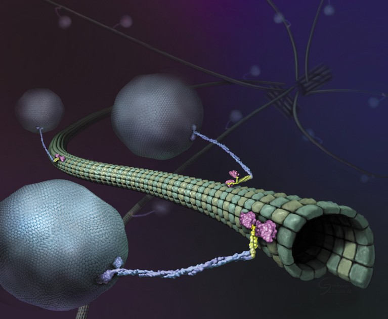
Illustration of molecular motors referred to as kinesins on a microtubule.Credit score: Graham Johnson, Ron Vale/HHMI
Virtually as quickly as there have been super-resolution microscopes, scientists pointed them in direction of molecular motors referred to as kinesins. These proteins, powered by the molecular gas ATP, drive essential processes together with cell division, cell signalling and intracellular transport by shuttling cargo alongside protein highways referred to as microtubules. Researchers have lengthy wished to know how these motors work, however to visualise them, scientists have needed to gradual them down or isolate them in simplified, in vitro methods.
Now, in papers printed concurrently in Science, two groups working independently have used a super-resolution instrument referred to as MINFLUX to review the motor in near-real time at physiologically related concentrations of ATP. The primary paper, led by MINFLUX’s inventor, Stefan Hell, who has a joint appointment on the Max Planck Institute (MPI) for Multidisciplinary Sciences in Göttingen and the MPI for Medical Analysis in Heidelberg, each in Germany, used a brand new instrument design to trace the protein in 3D, revealing particulars about its movement1. The second, led by biophysicist Jonas Ries on the European Molecular Biology Laboratory in Heidelberg, confirmed for the primary time that MINFLUX is able to monitoring kinesin even amid the bustle of dwelling cells2.
“This know-how requires plenty of various things to work, and it’s enjoyable to see all of these items coming collectively,” says Michelle Digman, a biomedical engineer on the College of California, Irvine, who develops imaging methods however was not concerned in both examine. “It appeared like a proof of idea to indicate that they’re in a position to monitor kinesin very exactly. And when you might have the reside cell system, that’s much more spectacular.”
Strolling the stroll
Researchers started understanding the fundamentals of kinesin’s movement shortly after the protein’s discovery in 1985, however the introduction of super-resolution instruments introduced a brand new degree of element. In 2004, researchers used a way referred to as FIONA (fluorescence imaging with one-nanometre accuracy) to indicate that kinesin, which seems to be like a tall, twisted stalk sporting outsized footwear, walks ‘hand-over-hand’ on its microtubule monitor, shifting its toes in a movement much like a baby’s palms as they traverse a set of monkey bars3. However, though FIONA offered distinctive spatial decision, the scientists needed to ration ATP to gradual the protein down sufficient to review it. Prior to now decade, researchers tagged kinesin with beads of germanium4 or gold5 to trace it, however these comparatively cumbersome tags go away doubt over how effectively the strategies recapitulate the protein’s full vary of movement.
When Hell and his group launched6 MINFLUX in 2016, he noticed it as an advance over its predecessor, stimulated emission depletion (STED) microscopy, for which Hell shared the 2014 Nobel Prize in Chemistry. STED makes use of a doughnut-shaped ‘depletion’ laser superimposed on an excitation laser to successfully shrink the world of fluorescence beneath the traditional diffraction restrict of sunshine (about 250 nanometres). MINFLUX, in contrast, makes use of a doughnut-shaped laser to create a degree of zero fluorescence depth at its centre. By shifting this laser, researchers can pinpoint a fluorescent molecule’s location at near-physiological speeds.

Sensible microscopes spot fleeting biology
Within the new examine1, printed in March, Hell’s group examined a model of MINFLUX that pulses linear lasers in two instructions within the focal aircraft in fast succession, localizing the protein by discovering the place the overlapping fluorescence intensities are lowest. By combining a number of measurements, the researchers had been in a position to produce tracks that present the place the molecule is shifting alongside the microtubule, like an app that maps out a runner’s path.
Though the brand new MINFLUX wasn’t far more efficient than the earlier iteration, Hell says, his group was ready to make use of it to infer a strolling velocity for kinesin of 550 nanometres per second and a stride size of 16 nanometres at ATP concentrations much like these present in dwelling cells. By tagging and monitoring completely different components of the protein, the group additionally confirmed that every stride contains two 8-nanometre substeps, and {that a} kinesin’s stalk rotates because it strikes, leading to a ahead movement that twirls barely to the proper. The authors additionally discovered that ATP is taken up when just one foot is sure to the microtubule, however is consumed when each toes are sure — resolving beforehand contradictory findings7,8.
Ries and his group approached kinesin movement from a unique perspective. They used a industrial MINFLUX instrument developed by Abberior, a Göttingen-based firm co-founded by Hell, to trace the protein in dwelling cells. The crowded and ever-changing mobile setting meant that the group ended up with fewer tracks, however they had been in a position to seize side-stepping, stalling and hops from one microtubule to a different — all efforts by the protein to avoid roadblocks that aren’t sometimes seen in purified samples. “In any other case, we see very a lot the identical issues: related strolling speeds, the identical step sizes,” Ries says. “And it was good to have the ability to measure these for the primary time within the dwelling cell.”
William Hancock, a biomedical engineer at Pennsylvania State College in State Faculty who research kinesin however was not concerned in both examine, calls the research “fairly unbelievable”. The outcomes, he notes, largely gel with previous work, nearly all of which had been executed in vitro utilizing purified motor proteins. “We get our very particular solutions there, nevertheless it’s good to have the ability to extrapolate again to the within of cells. It’s actually a tour de drive.”
Motoring forwards
That stated, it’s clear to Hell that MINFLUX “isn’t but on the restrict of its efficiency”. Having achieved subnanometre spatial decision, one space the place MINFLUX can nonetheless enhance, he says, is in its temporal efficiency. Each teams labored with ATP concentrations of as much as one millimolar, however the common cell incorporates greater than 4 instances as a lot.
Reaching real-time temporal decision beneath these situations would require higher monitoring methods, says Luke Lavis, an natural chemist on the Howard Hughes Medical Institute’s Janelia Analysis Campus in Ashburn, Virginia, who developed the dyes utilized in Ries’s examine. “This revolution in super-resolution imaging brings into focus, if I can say that, the truth that plenty of the labelling and attachment methods we’ve used are massive,” he says. “The dream for all of us is having the ability to connect a small-molecule fluorophore to a particular place on a protein the place the linker is simply a few atoms.” New instruments similar to genetic code growth, wherein artificial amino acids are engineered into proteins to facilitate labelling, are shifting on this course, he provides, however “they’re removed from being turn-key”.

NatureTech hub
Such advances may open a number of analysis avenues, particularly if they permit researchers to trace a number of proteins, or a number of websites in proteins, without delay. This yr, researchers launched a instrument referred to as RESI (decision enhancement by sequential imaging), which goals to just do that9. RESI can label adjoining copies of the identical goal molecule with completely different tags, permitting scientists to tell apart between molecules which might be lower than one nanometre aside. Though RESI presently works solely on fastened or stationary molecules, combining its findings with MINFLUX monitoring information on non-fixed copies may yield complementary findings a couple of protein’s association and movement.
Ries is focused on finding out different motor proteins, and in a supplementary experiment shared in his paper2, utilized MINFLUX to the muscle-contracting protein, myosin. In 2019, Lavis co-founded an organization to make use of single molecule monitoring for drug improvement. Different scientists have steered all the pieces from transcription elements and DNA-binding proteins to cohesin and different molecular motors as targets for future examine.
However Haley Marks, a biomedical engineer and venture scientist on the California NanoSystems Institute on the College of California, Los Angeles, says it would take time earlier than MINFLUX turns into a very accessible and inexpensive “plug-and-play” system. Within the meantime, she suggests, MINFLUX may benefit the broader group by contributing to digital super-resolution instruments. These artificial-intelligence algorithms prepare on giant collections of microscopy pictures to discover ways to create super-resolution pictures from typical microscopy ones. The ensuing information, Marks says, “are far more accessible by individuals who can’t afford a multimillion-dollar microscope”.
[ad_2]